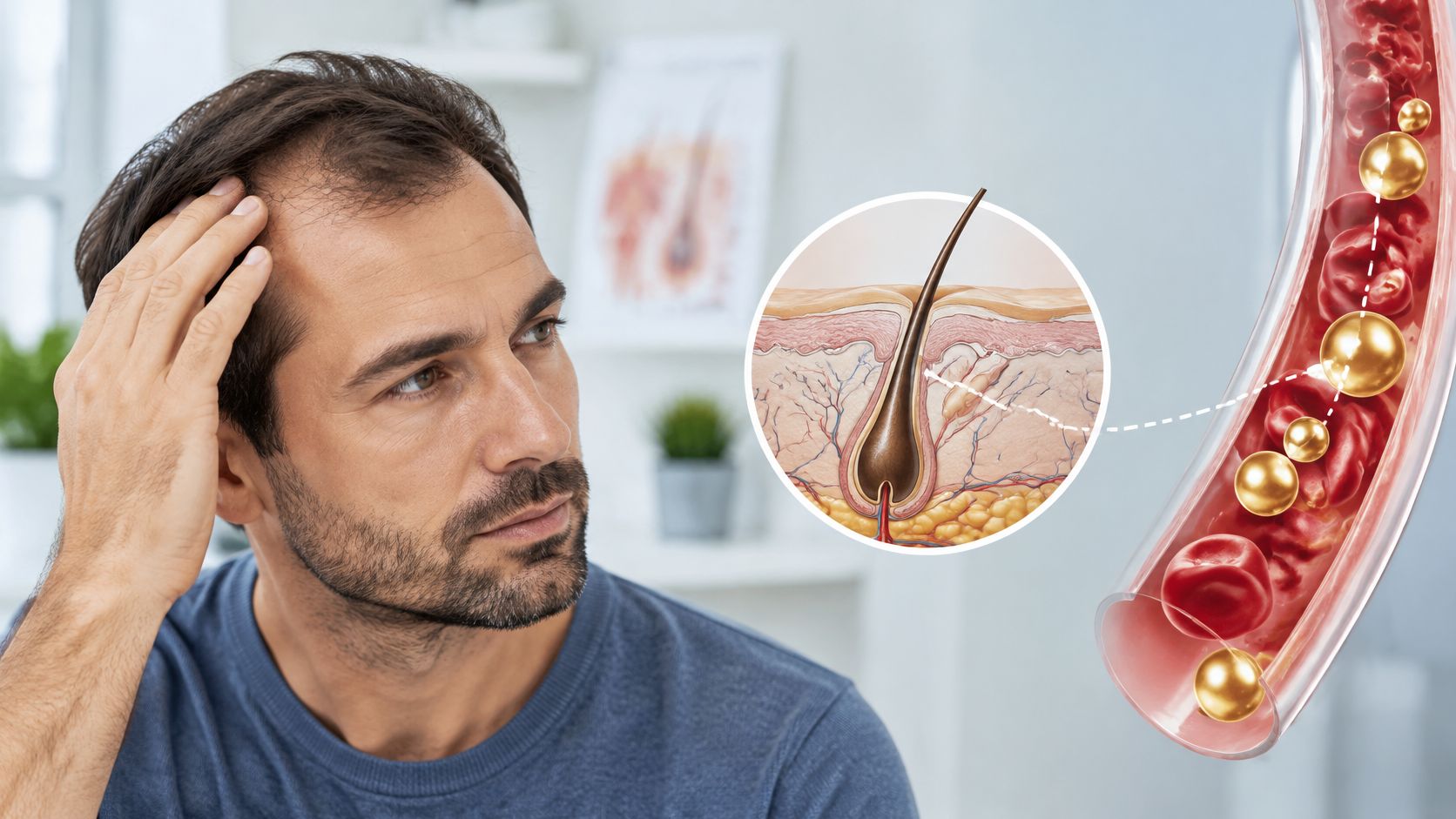
Crown of Your Head: Thinning and Care Guide
Learn what the crown of your head is, how vertex hair loss shows, which scalp signs need care, and how to track change.
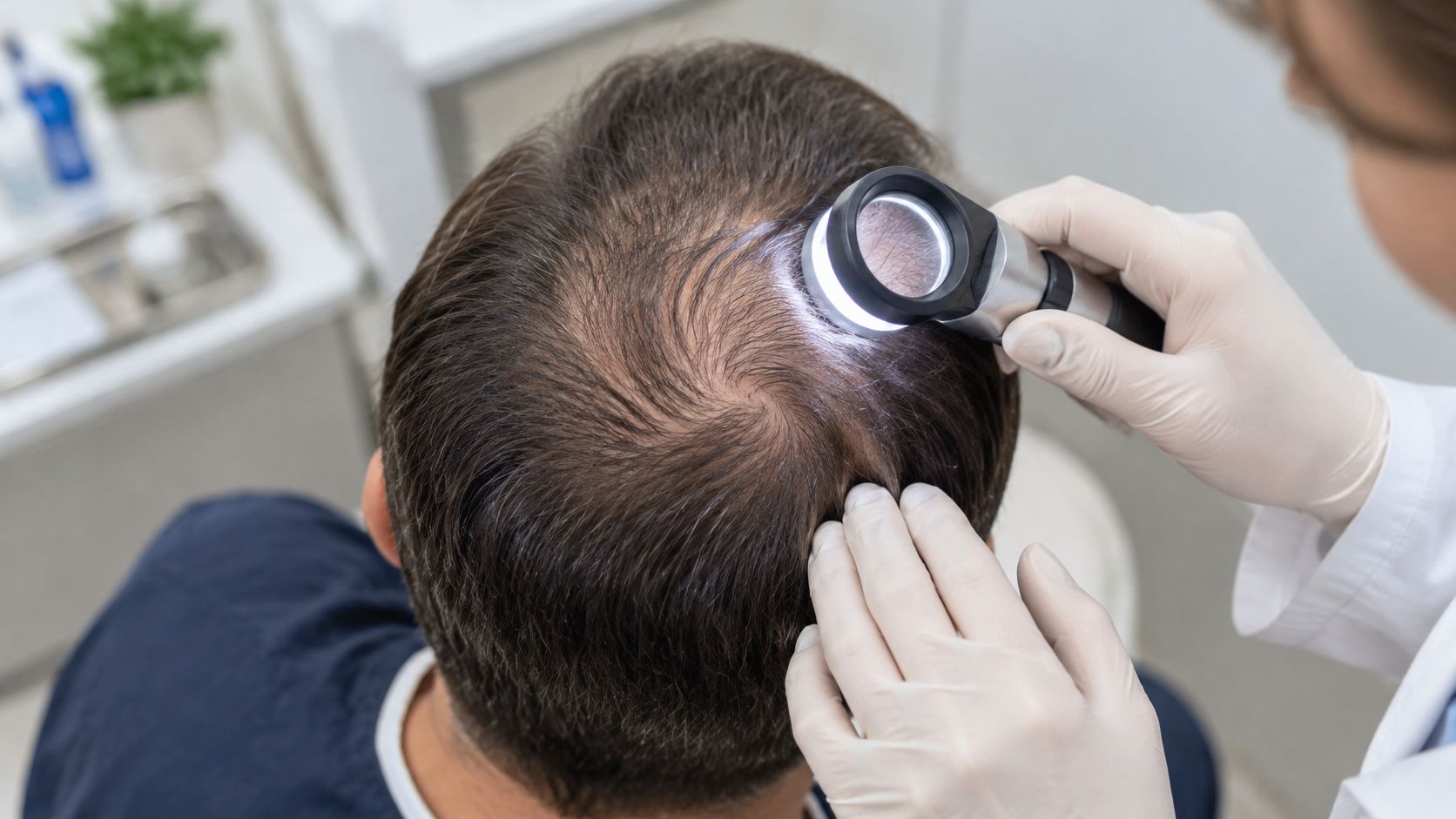
A wider crown is not a diagnosis. Treating it as one is how people spend months on a product that never matched the cause.
The crown of your head is the vertex, the top-center scalp around the natural hair whorl. Hereditary thinning often shows there, but a visible crown alone does not confirm androgenetic alopecia. Gradual pattern loss, temporary shedding, and inflammatory or scarring scalp disease can all change the same area. They call for different decisions.
Crown-specific triage begins with pattern, symptoms, and progression. It separates ordinary vertex thinning from scalp disease that needs prompt review. A casual phone photo rarely settles the question, because the whorl is hard to see, lighting hides early change, and angle can exaggerate or mask density. A consistent clinical view does.
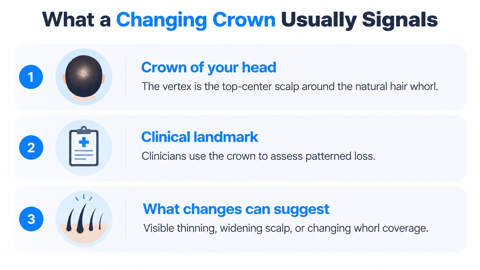
What a Changing Crown Usually Signals
The crown, or vertex, is the top-center scalp surrounding the natural hair whorl. Clinicians use this region as a landmark for patterned hair loss, but its location doesn't identify the cause by itself.
Crown findings and how quickly to seek care
| Finding | Pattern it may fit | Suggested urgency |
|---|---|---|
| Gradual widening with temple or mid-frontal recession | Male androgenetic alopecia | Routine clinical review |
| Diffuse crown thinning with preserved frontal hairline | Female pattern hair loss | Routine clinical review |
| Round smooth patches or broken hairs | Possible alopecia areata | Prompt evaluation |
| Itch, burning, tenderness, scale, or pustules | Inflammatory or infectious scalp disease | Prompt evaluation |
| Shiny skin and lost follicular openings at the crown | Possible scarring alopecia, including CCCA | Urgent dermatologic evaluation |
| Sudden heavy shed after illness, stress, or a medication change | Possible telogen effluvium | Review if shedding persists or the pattern changes |
The Merck Manual's clinical guide to alopecia distinguishes patterned loss from patchy, inflammatory, infectious, and scarring causes. These categories help establish urgency, but the table isn't a substitute for examining the scalp.
Gradual changes and warning signs
Slowly reduced coverage accompanied by temple recession often fits male androgenetic alopecia. In women, broader thinning across the crown with preservation of the frontal hairline may fit female pattern hair loss. Neither appearance is diagnostic without considering the history and scalp findings.
Round, smooth patches raise a different question, including possible alopecia areata. Broken hairs also warrant evaluation because they can occur with infection, mechanical damage, or other disorders. A clinician considers the skin beneath the hair rather than assigning a diagnosis from the outline of the patch.
Itching, burning, tenderness, scale, and pustules shouldn't be dismissed as ordinary genetic thinning. These symptoms can indicate an inflammatory or infectious process that needs targeted treatment rather than an empiric hair-growth routine.
Why suspected scarring changes the priority
Central centrifugal cicatricial alopecia, abbreviated CCCA, often begins at the crown and disproportionately affects middle-aged Black women. Smooth, shiny areas with absent follicular openings are concerning because scarring disease can permanently destroy follicles. Delayed assessment can therefore reduce the opportunity to preserve remaining hair.
A symptom-free photograph also requires careful interpretation. Hair clumping and scalp visibility change with moisture, as explained in this guide to hair that looks thin when wet. An appearance change caused by wetness isn't equivalent to documented follicle loss.
A questionnaire or photo set cannot exclude scarring alopecia. Uncertain, symptomatic, or rapidly progressing cases may require trichoscopy and sometimes a scalp biopsy.
Trichoscopy provides a magnified examination of hair shafts, follicular openings, and scalp skin. A biopsy may be necessary when the diagnosis remains uncertain or scarring is suspected. In those circumstances, arranging an examination takes priority over comparing cosmetic products.
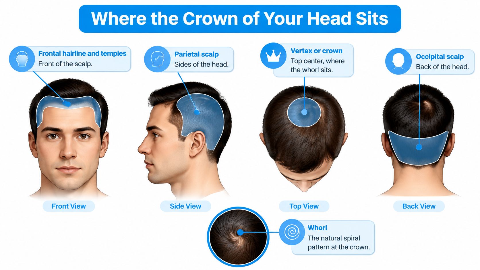
Where the Crown of Your Head Sits
A simple scalp map starts at the forehead and moves backward. The frontal hairline and temples occupy the front, the parietal scalp lies along the sides, the vertex sits near the top center, and the occipital scalp covers the back of the head.
Someone examining the crown with mirrors may notice a small area of visible skin where hairs spiral outward. That whorl is a normal growth pattern. Because the strands point in different directions at its center, the scalp can look exposed without a meaningful change in hair counts.
From a normal follicular unit to miniaturization
A follicular unit is a small grouping of follicles emerging together through the scalp. Units normally produce approximately two to five hairs, while androgen-affected units progressively lose terminal-hair coverage, as described in this clinical review of androgenetic alopecia.
Terminal hairs are the thicker, more pigmented strands that provide most visible coverage. During miniaturization, susceptible follicles produce progressively shorter, finer, vellus-like hairs. The scalp may become easier to see before a distinct bald patch develops.
The biochemical pathway involves dihydrotestosterone, or DHT, and androgen-sensitive cells in the follicle's dermal papilla. Androgen signaling shortens anagen, the active growth phase, and contributes to progressive miniaturization. Vertex follicles are generally more sensitive to this signaling than follicles in the occipital region.
That regional difference explains why the back of the scalp is useful as a comparison and is often considered for donor hair in transplantation. It doesn't mean every occipital follicle is unaffected or every visible crown is diseased.
What magnification can reveal
A clinician examining the crown looks for more than the size of the whorl:
- Uneven shaft thickness: Greater hair-diameter diversity can indicate that some follicles are miniaturizing while neighboring follicles still produce thicker strands.
- More fine hairs: A higher proportion of thin shafts can reduce coverage even when many hairs remain visible.
- Reduced terminal-hair grouping: Fewer thick hairs emerging from each unit can make the scalp more apparent.
For example, a crown may look reasonably covered when brushed forward but sparse when the hair separates around the spiral. Comparing that region with the occipital scalp, ideally through clinical examination, is more informative than judging the whorl in isolation.
How Often the Vertex Thins With Age
Vertex prevalence, site frequency, and density
| Measure | Group | Figure |
|---|---|---|
| Androgenetic alopecia prevalence | Men by age 50 | About 30% to 50% |
| Vertex or complete baldness | Men aged 40–55 | 31% |
| Vertex or complete baldness | Men aged 65–69 | 53% |
Age alone does not explain a thinning crown, but age bands make the pattern easier to read. The National Institutes of Health's Endotext reference reports vertex or complete baldness in 31% of men aged 40–55 and 53% of men aged 65–69. Those figures sit inside a broader picture of androgenetic alopecia that is common by midlife, yet they still describe groups, not one person's scalp.
| Measure | Group | Figure |
|---|---|---|
| Most frequent hair-loss site | 300 people with scalp hair loss | Vertex, 97 cases (32.33%) |
| Next most frequent sites | Same 300-person series | Parietal 19.33%; occipital 17.33% |
| Vertex hair density | Female-pattern loss vs controls | 112.05 vs 140.21 hairs/cm² |
| Vertex shaft diameter | Female-pattern loss vs controls | 54.23 vs 67.38 micrometers |
The published quantitative assessment of female-pattern hair loss shifts from age bands to clinical detail. In a 300-person series, the vertex was the most frequent site, with 97 cases (32.33%), ahead of the parietal region (19.33%) and occipital region (17.33%). A separate comparison of 38 participants with female-pattern loss and 35 controls found lower vertex density, 112.05 versus 140.21 hairs/cm², and finer shaft diameter, 54.23 versus 67.38 micrometers, roughly 20% lower on each measure.
What the measurements mean for coverage
Density counts how many hairs occupy an area. Shaft diameter describes how thick each strand is. A drop in either reduces visual coverage, but the two are separate measurements. They do not add into a single percentage of visible baldness, and a person may notice less fullness without collecting much more hair in the shower.
Density and shaft diameter work like the number of trees and the width of their trunks. Both affect how much ground they cover. The crown is a useful but tricky monitoring site. The whorl can hide early change because hair lies in different directions and lighting alters contrast. A casual photo may flatten the pattern or crop out the temples and frontal hairline, so it can suggest a problem that is not there or miss one that is. Clinical examination looks at the scalp surface, the distribution beyond the whorl, and symptoms that point away from ordinary patterned loss.
The prevalence numbers answer a narrow question: how often vertex involvement appears in studied groups. They do not distinguish common androgenetic vertex thinning from an inflammatory or scarring process in an individual. A clinical series of people already seeking care describes site frequency within that sample, not crown-loss prevalence in the general population. A comparison between participants with female-pattern hair loss and controls shows group differences, not a home diagnostic cutoff. No phone photo can reliably measure hairs per square centimeter or shaft diameter.
Family history, pattern, and symptoms help a clinician interpret those measurements. Age raises the likelihood of patterned loss, but it does not confirm the cause. A clinician can separate the possibilities through a scalp examination.
How to Track a Crown You Cannot See
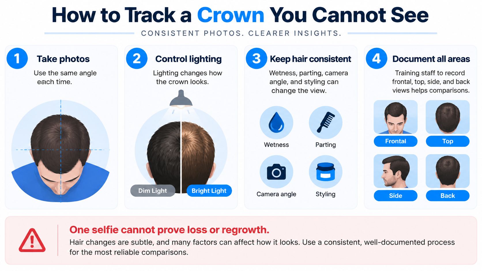
Informal crown photographs can differ because of lighting, wetness, part placement, camera angle, and styling. A bright overhead bulb can expose scalp between strands that appears well covered in softer light. A tilted camera can make the same whorl look broader or narrower.
Documentation quality is something clinicians can improve deliberately. A clinical scalp-photography study found that staff training significantly improved hair-loss photograph quality, with p < 0.005. That finding supports consistent technique, not the assumption that any photograph can establish a diagnosis.
A repeatable home photography routine
- Fix the camera position. Use the same phone and camera setting, with a tripod or another stable arrangement that preserves distance. A helper can make a top-down view easier to reproduce.
- Photograph dry, unstyled hair under the same overhead light. Avoid comparing a wet post-shower image with a dry, freshly styled image.
- Record several views. Include a true top-down crown photograph, a frontal view, and views of both temples. A crown-only image can miss a changing pattern elsewhere.
- Keep the part in the same place and arrange the surrounding hair consistently. Don't pull strands apart more forcefully in one session than another.
- Choose a recurring interval with the treating clinician. For a hypothetical home schedule, photographs every three months may be easier to compare than daily images, but symptoms or rapid changes require earlier review.
- Save dated originals and compare them side by side. Avoid filters, contrast adjustments, or selective cropping that changes how much scalp appears visible.
What the images cannot prove
A naturally wide whorl or temporary shedding can resemble progressive pattern loss, while slow miniaturization may be missed in casual selfies. A single image cannot establish loss or regrowth, and a more favorable hairstyle isn't evidence that follicles have changed.
Baseline photographs are most useful before starting treatment or changing a regimen. Notes should record relevant context, such as a recent illness, medication change, haircut, or new scalp symptom, without treating any one event as the confirmed cause.
Consistent images can improve remote triage and help a clinician decide whether an in-person examination is needed. They cannot independently rule out scarring alopecia or other scalp disease. The appropriate response to concerning symptoms is clinical assessment, not increasingly frequent photography.
Medicines That Can Slow Vertex Loss
Medication selection follows the diagnosis, not the photograph. A wide whorl can look alarming in a phone snapshot and still be ordinary scalp anatomy, so the first question is whether the crown shows patterned follicular miniaturization at all. When androgenetic alopecia is the working diagnosis, treatment aims to preserve hair and support growth. Holding present density is a realistic outcome; rebuilding the original crown coverage is not.
The American Academy of Dermatology's male-pattern hair-loss treatment guide reports that finasteride slows further loss in approximately 80% to 90% of men who take it, with improvement potentially noticeable after about four months, while minoxidil results commonly require six to twelve months. Those intervals are why a two-week trial teaches nothing about whether a medicine is working.
Vertex hair therapies, actions, and typical timelines
| Option | Main action | When change may show | Important limit |
|---|---|---|---|
| Topical minoxidil | Supports anagen; linked to potassium-channel opening | Six to twelve months | Continued use needed to hold any gain |
| Oral finasteride | Inhibits type II 5-alpha-reductase and lowers DHT | About four months | Prescription only; not for anyone who is pregnant or may become pregnant |
| Dutasteride | Inhibits type I and type II 5-alpha-reductase | Assessed individually; no set public timeline | Needs assessment of individual risk and tolerability |
| Combined or compounded plans | Selected mechanisms used together | Assessed according to the prescribed plan | Selected only after review of the whole history |
Different pathways, different prescribing decisions
Finasteride reduces the conversion of testosterone to DHT by inhibiting type II 5-alpha-reductase. Dutasteride inhibits both type I and type II forms of that enzyme, a broader block along the same pathway. Broader inhibition does not establish that dutasteride suits a particular patient; the comparison of dutasteride versus finasteride for hair loss turns on individual risk, tolerability, and the goal the clinician sets.
Minoxidil supports follicular activity through mechanisms associated with potassium-channel opening and anagen support, a route separate from DHT suppression. That separation is the practical reason these medicines are not interchangeable, and why a switch belongs in a prescribing conversation rather than a product comparison.
The American Academy of Dermatology's diagnosis and treatment guide identifies topical minoxidil as FDA-approved for hair loss in men and women, while oral finasteride is FDA-approved for male pattern hair loss. For women with diffuse crown thinning and a preserved frontal hairline, topical minoxidil is a usual starting consideration after diagnosis.
Safety and maintenance matter
Finasteride may be prescribed selectively to women under medical supervision, with careful attention to pregnancy potential and contraception. It isn't appropriate for anyone who is pregnant or may become pregnant because of fetal risk. Hormonal evaluation is considered separately when the history or examination suggests a systemic cause.
- Route matters: Topical and oral therapies carry different safety considerations, including cardiovascular history, scalp disease, and medication interactions.
- Maintenance is ongoing: Benefits generally require continued treatment; stopping can allow the underlying process to resume.
- Combinations require review: A compounded or combined formula is not automatically preferable to a single medication.
Prescribing and compounding sit with a licensed U.S. clinician after an individualized intake, because a crown photograph cannot confirm either the diagnosis or the dose.
Everyday Care That Does Not Diagnose Loss
A wide crown whorl does not automatically mean genetic thinning. Everyday care can protect scalp comfort and reduce avoidable hair damage, but it cannot distinguish common vertex pattern loss from an inflammatory or scarring condition. That distinction depends on the pattern and scalp findings, not on how a hairstyle looks after washing.
A gentler routine may help strands separate and lie more evenly over the crown. This changes the appearance of coverage, much like arranging overlapping fibers, but does not show whether follicular density has changed.

Supportive habits without regrowth claims
- Clean the scalp gently. Set a wash schedule that removes oil, sweat, and product residue. Massage shampoo with your fingertips rather than scraping the whorl with your nails.
- Reduce sustained pulling. Very tight styles can place unnecessary tension on hair. Adjust a style that causes soreness or persistent pulling.
- Manage product buildup. Heavy styling products can clump strands and make crown coverage harder to judge. If irritation persists, stop the suspected product and arrange an assessment.
- Limit repeated high heat. Use lower heat when practical, and avoid passing a hot tool over the same crown strands repeatedly.
- Protect exposed scalp. A hat or suitable sunscreen can help protect visible vertex skin from sunburn. Hair coverage may not fully protect the scalp.
- Leave scale and prescriptions alone. Picking can injure irritated skin. Another person's prescription may not suit your scalp condition.
Choose washing frequency based on scalp and styling needs, not a rigid belief that less washing preserves follicles. Shampoo can loosen hairs that were already ready to shed, so their appearance during washing does not identify the cause of hair loss.
Shedding and supplements are easy to misread
For minoxidil-related shedding, see the treatment guidance above. A change in shedding should be interpreted in context, especially if it is marked, persistent, or accompanied by new scalp symptoms.
Vitamins and cosmetic oils also have limits. A clinically identified nutrient deficiency may need correction, while supplements or oils should not be expected to reverse androgen-driven miniaturization. Applying several products can also make it harder to identify what is irritating the scalp.
Family history and age provide context, but the pattern across the temples, mid-scalp, and crown also matters. A relative with a similar-looking whorl does not establish the cause in another person. Casual crown photos can mislead because lighting, hair direction, and the position of the whorl change how much scalp shows.
Keep comfortable, tolerable habits, and pay attention to symptoms rather than relying on cosmetic changes. Scalp inflammation or consistent photographic decline calls for a closer review, since routine care cannot sort those findings into a diagnosis.
When to Request a Clinical Hair Review
A changing crown can reflect common vertex-pattern loss, but inflammation or scarring calls for a different evaluation. Request clinical review for any prompt or urgent finding in the triage table, persistent shedding after illness or a medication change, or a sustained density decline across repeated photo comparisons. A wide whorl can also be difficult to interpret from casual pictures, so uncertainty is a reasonable reason to seek assessment. Younger adults should not assume age rules out clinically significant loss; see this guide to hair loss in young men.
Prepare a useful intake
- History: Age, family history, when changes began, and recent illness or medication changes.
- Safety information: Pregnancy status, reproductive plans, and a complete list of medications and supplements.
- Documentation: Standardized crown, frontal, and temporal photographs, along with descriptions of discomfort or visible scalp changes.
Vials + Vitals' hair health area offers three oral hair options, each with its listed ingredients. A clinician considers the individual's assessment when selecting whether an option is appropriate and which formulation to use.
Availability depends on state rules, and a consultation does not guarantee a prescription. If treatment is not appropriate, the provider will document that recommendation. Suspected scarring may require in-person trichoscopy or biopsy, rather than remote prescribing.
Informational purposes only. A licensed provider evaluates compounded medications for individual suitability, and state-licensed partner compounding pharmacies fulfill them.
Vials + Vitals offers physician-directed telehealth assessment for adults considering medically supervised hair treatment. Visit Vials + Vitals to begin an individualized intake with your scalp history, medication list, and consistent photographs.