Visceral Fat Check Guide: How to Measure and Read Results
Learn how to run a visceral fat check with waist measurements, ratios, and imaging options, and how to read results with a licensed clinician.

The 2025 VA/DoD obesity guidance found insufficient evidence to recommend for or against a particular adiposity measure for managing clinical outcomes, a useful caution against treating any visceral fat score as the definitive answer (VA/DoD obesity guideline presentation). A more detailed measurement doesn't automatically lead to a better treatment decision. What matters is whether the result helps a clinician assess risk or change care.
A visceral fat check works best as a repeatable protocol, not a search for a single ideal number. Visceral fat surrounds abdominal organs; subcutaneous fat lies beneath the skin. Home measurements can help screen for abdominal adiposity, but they can't show exactly how much fat lies in each compartment.
Visceral Fat Check Methods Compared
| Method | What It Measures | Radiation | Access and Cost | Best Use |
|---|---|---|---|---|
| Waist tape | Abdominal circumference, includes subcutaneous fat | None | Low, at home | Routine screening and trend tracking |
| BMI | Weight relative to height, no fat distribution | None | Low, at home | Reference point only |
| Smart scale BIA | Estimated body water and fat, device-specific | None | Low, at home | Trends under identical conditions |
| DXA | Whole-body fat and regional estimates, no direct visceral split | Low | Moderate, clinic-based | Standardized body composition |
| Ultrasound | Abdominal fat depth at a set site | None | Moderate, operator-dependent | Practical intermediate measure |
| CT | Visceral and subcutaneous area or volume | Ionizing | High, specialized | Precise compartment measurement when indicated |
| MRI | Visceral and subcutaneous area or volume | None | High, least widely available | Precise measurement without radiation |
These methods move broadly from inexpensive screening toward specialized compartment measurement. They aren't a universal ranking: ultrasound quality relies on operator technique, and access can influence which test is practical. No row represents a stand-alone diagnosis.
What the instruments actually detect
Bioelectrical impedance analysis, or BIA, uses electrical impedance to estimate body water and infer body composition. A smart scale doesn't image abdominal organs or directly weigh visceral fat. Its displayed visceral fat level is a device-specific estimate.
A review in Clinical Epidemiology and Global Health00149-5/fulltext) characterizes BIA as the least accurate common surrogate for visceral adiposity and reports DXA correlations of r = 0.902 with MRI-measured visceral fat volume and r = 0.83 with CT-measured visceral fat area. Strong correlation means measurements tend to move together across a group, not that their absolute results are interchangeable.
DXA uses low-dose X-rays to estimate tissue composition across a two-dimensional image. Its abdominal software estimates visceral fat rather than directly separating deep and superficial fat compartments. CT and MRI can distinguish those compartments more directly, while ultrasound measures depth at a defined location.
Matching the method to the situation
Routine screening without concerning symptoms generally begins with waist circumference and BMI. For readers comparing weight, fat percentage, and abdominal measurements, the distinction between male body fat and fat distribution helps explain why these measures answer different questions.
Home or telehealth monitoring can incorporate BIA when the record includes the device model, electrode configuration, time of day, fasting status, fluid intake, recent exercise, bladder status, and menstrual or edema-related changes. Proprietary visceral scores shouldn't be compared across manufacturers.
Clinician-ordered DXA or ultrasound may add useful information when a more structured assessment is needed. Ultrasound requires controlled positioning, breathing, probe orientation, and pressure. CT or MRI is most defensible when the result would change management, rather than merely satisfy curiosity.
Practical rule: A new device creates a new baseline unless a clinician establishes that the results can be compared.
The most common interpretation error is switching methods across visits. A different anatomical slice, software version, operator, or hydration state can create apparent change without corresponding fat loss. Vials + Vitals content is educational only and isn't medical advice.
How to Measure Your Waist Correctly
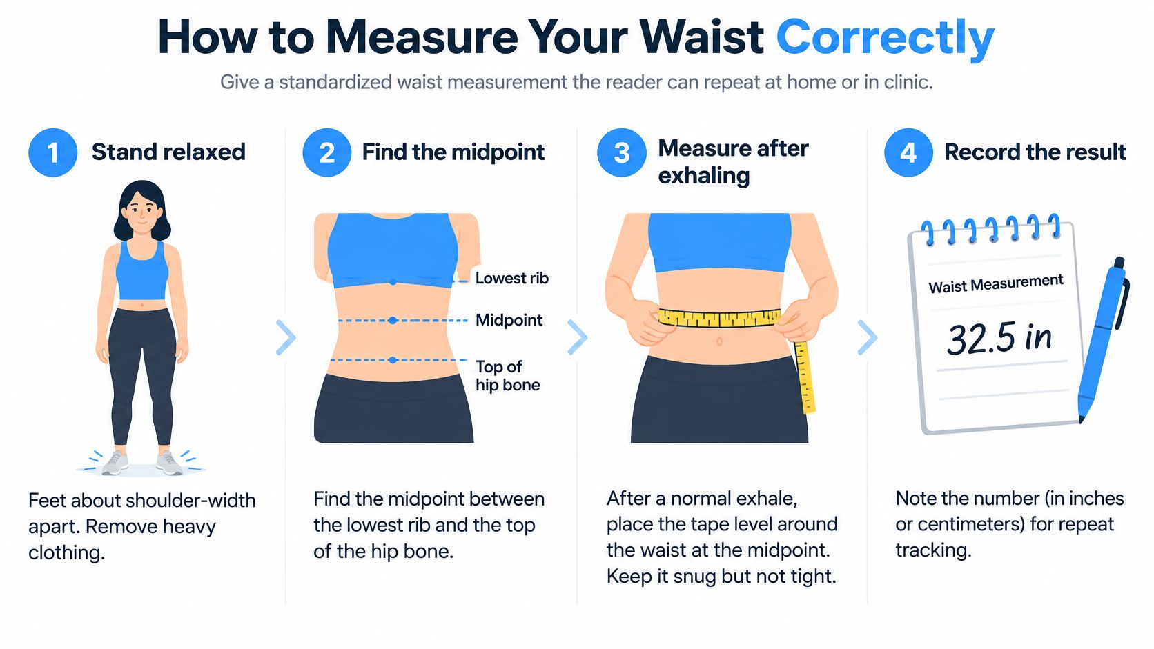
A useful waist measurement begins with a nonstretch measuring tape, a consistent anatomical landmark, and relaxed breathing. The number should describe the abdomen, not how tightly the tape was pulled.
The illustration summarizes the basic positioning and recording process.
A repeatable home technique
Stand relaxed. The person being measured stands with feet approximately shoulder-width apart and removes heavy clothing from the waist. A relaxed stance limits posture-related differences, while measuring bare skin avoids adding fabric thickness.
Find the midpoint. Locate the lowest rib and the top of the hip bone, also called the iliac crest, then identify the midpoint between them. Using a reproducible landmark is more reliable than choosing the narrowest-looking part of the waist or assuming the navel is the correct site.
Position the tape. Wrap the nonstretch tape around the bare abdomen, level with the floor and snug without indenting the skin. A mirror or another person's assistance can help identify a tape that slopes downward across the back.
Read after a normal exhalation. The abdomen stays relaxed while the person breathes out normally, without forcefully emptying the lungs or drawing in the stomach. Breath-holding and deliberate bracing make later comparisons less dependable.
Repeat and average. Remove and reposition the tape for repeat readings, then record their average. Repositioning helps reveal placement errors that would remain hidden if the tape never moved.
Record the conditions. Note the date, time, clothing, and whether the measurement followed a meal or exercise. A large meal can temporarily increase abdominal circumference, while excessive tape tension can artificially reduce it.
The midpoint, standing posture, nonstretch tape, normal exhalation, and repeated measurement approach align with the British Journal of Nutrition validation study of abdominal adiposity measurements. Waist circumference remains an indirect proxy because it includes superficial as well as deeper abdominal tissues.
What belongs in the measurement record
A usable entry contains a dated average, body weight, measurement site, and conditions. Recording the individual readings also helps a clinician assess whether the average came from closely matched measurements or inconsistent technique.
The same tape and time of day make follow-up easier to interpret. If a clinic uses a different waist landmark, that difference should be documented rather than combining the readings.
A carefully recorded waist measurement is more useful than a precise-looking number taken at an unknown location.
Reading Waist and Ratio Results
A waist cutoff is a screening flag, not a diagnosis. The thresholds below are commonly cited reference points, but interpretation depends on the measurement method and the person's context.
| Measure | Commonly cited higher-risk cutoff | Key caveat |
|---|---|---|
| Waist circumference, men | 102 cm (40 in) or more | Cutoffs vary by ancestry |
| Waist circumference, women | 88 cm (35 in) or more | Use a consistent measurement method |
| Waist-to-hip ratio, men | Above 0.90 | Compares waist size with hip size |
| Waist-to-hip ratio, women | Above 0.85 | Compares waist size with hip size |
| Waist-to-height ratio, adults | 0.5 or more | Relates waist size to height |
A result below a common cutoff does not rule out metabolic disease, and a result above it does not establish a diagnosis. Clinicians may use different thresholds based on ancestry, sex-related risk patterns, and the guideline relevant to the patient.
What each ratio adds
Waist-to-height ratio divides waist circumference by height, using the same units for both. It places abdominal size in context: a given waist measurement means something different for a shorter adult than for a taller one.
Waist-to-hip ratio divides waist circumference by hip circumference, usually measured around the widest part of the buttocks. It describes waist size relative to hip size. Since either measurement can change, a lower ratio does not necessarily mean abdominal fat has decreased.
Neither ratio measures the visceral-to-subcutaneous fat ratio. That measure compares deep abdominal fat with fat beneath the skin, while waist-based ratios compare external body dimensions.
Why fat distribution matters
A 2024 systematic review of visceral-to-subcutaneous abdominal fat ratios examined 27 studies and found that higher ratios were generally associated with cardiovascular disease risk. The evidence was primarily observational, so it supports risk assessment but does not show that a particular ratio causes an event.
Adults with similar BMI can carry weight differently and have different clinical risk profiles. BMI relates weight to height, but it does not describe where weight is carried. Waist and ratio results therefore add context to an assessment that also considers blood pressure, glucose regulation, lipid results, medications, and medical history.
The measurement protocol matters here: a ratio calculated from a waist taken at one landmark cannot be reliably compared with a later ratio based on a different landmark. Consistent methods make the numbers easier to interpret, while clinical context determines what they mean.
Tracking Change Without Overreading Results
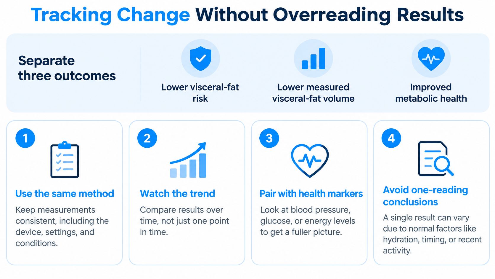
Lower visceral-fat risk, lower measured visceral-fat volume, and improved metabolic health are different outcomes. They may occur together, but a change in one doesn't prove an equal change in the others. A smaller waist suggests a different external measurement; it doesn't quantify the amount of deep fat lost.
The tracking checklist keeps those outcomes separate while organizing follow-up.
A follow-up checklist
- Preserve the baseline protocol. Follow-up uses the same method, device, anatomical site, and preparation. If equipment changes, the record should identify the change rather than imply uninterrupted comparability.
- Assess a trend over weeks. Measurements several weeks apart under comparable conditions are more useful than repeated reactions to daily fluctuations. The appropriate interval depends on the clinical question and treatment plan.
- Keep related markers together. Standardized waist measurements, weight, blood pressure, glucose or HbA1c, triglycerides, HDL, relevant liver indicators, and symptoms belong in the same longitudinal record.
- Verify a surprising result. An unexpected change warrants a technique check and confirmation with another relevant marker before treatment is adjusted.
For people receiving GLP-1 or GIP-based treatment, total weight loss can include fat-free mass. The scale therefore can't establish which tissue compartment changed, and abdominal measurements need not improve in exact proportion to body weight.
Visible change is assessed over months rather than days, and a response isn't guaranteed. Understanding how long weight loss may take to become noticeable can help separate reasonable follow-up expectations from daily checking.
Better automation doesn't settle every clinical question
A 2025 MRI study of automated volumetric measurements reported agreement with manual reference measurements with intraclass correlation coefficients of at least 0.96 and coefficients of variation no higher than 3.8%. Those results support repeatability within the evaluated measurement approach, not universal equivalence across scanners or methods.
More repeatable imaging can improve measurement quality without establishing that every patient needs imaging.
A practical decision rule is whether a scan would resolve uncertainty that could change care. When clinical markers already support a clear plan, another anatomical number may add little.
Deciding Your Next Step With a Licensed Provider
A measurement trend becomes useful in a clinical conversation when it connects to your priorities and treatment experience. Refer to the measurement record you have already established, rather than rebuilding it from memory. A provider can then discuss what the pattern may mean alongside relevant clinical context.
At Vials + Vitals, the first step is an online medical intake covering health history, symptoms, and treatment goals. An independent, licensed healthcare provider reviews the information against the patient's chart and may request more details. Consultations are conducted by licensed U.S. healthcare providers.

How treatment decisions differ from measurement decisions
A change in body weight does not, by itself, show whether visceral fat changed. Semaglutide and tirzepatide act on appetite-related pathways, and treatment-related weight loss may involve different tissue compartments. That is why waist measurements and other relevant indicators need consistent conditions and clinical interpretation, rather than being treated as direct substitutes for a measure of visceral fat.
Medication decisions are separate from measurement decisions. A licensed provider considers the specific prescription, indication, tolerability, and patient risk profile. Follow-up may address symptoms, adherence, and metabolic markers.
Nutrition, appropriate physical activity, sleep, and sustainable routines are also part of improving metabolic health. A “metabolic reset” is not a defined measurement endpoint or a promised treatment outcome.
Vials + Vitals provides marketing, management, and patient-intake access, not medical care or prescribing. An independent physician may approve, modify, defer pending labs, or decline a request. A prescription or outcome is never promised.
What belongs in the consultation record
Bring the priorities that measurements alone cannot capture:
- Patient priorities: Symptoms, treatment goals, and what changes matter most to you.
- Medication context: Current medications and supplements that may affect treatment decisions.
- Tolerability concerns: Previous side effects, concerns about treatment, and any barriers to following a plan.
The clinician can interpret these details alongside the measurement record and clinical context already gathered. Vials + Vitals content is educational only and is not medical advice. Health information belongs on the program's dedicated clinical intake page, not on marketing pages.
Adults seeking an individualized assessment can visit Vials + Vitals and complete the online intake for independent licensed-clinician review.