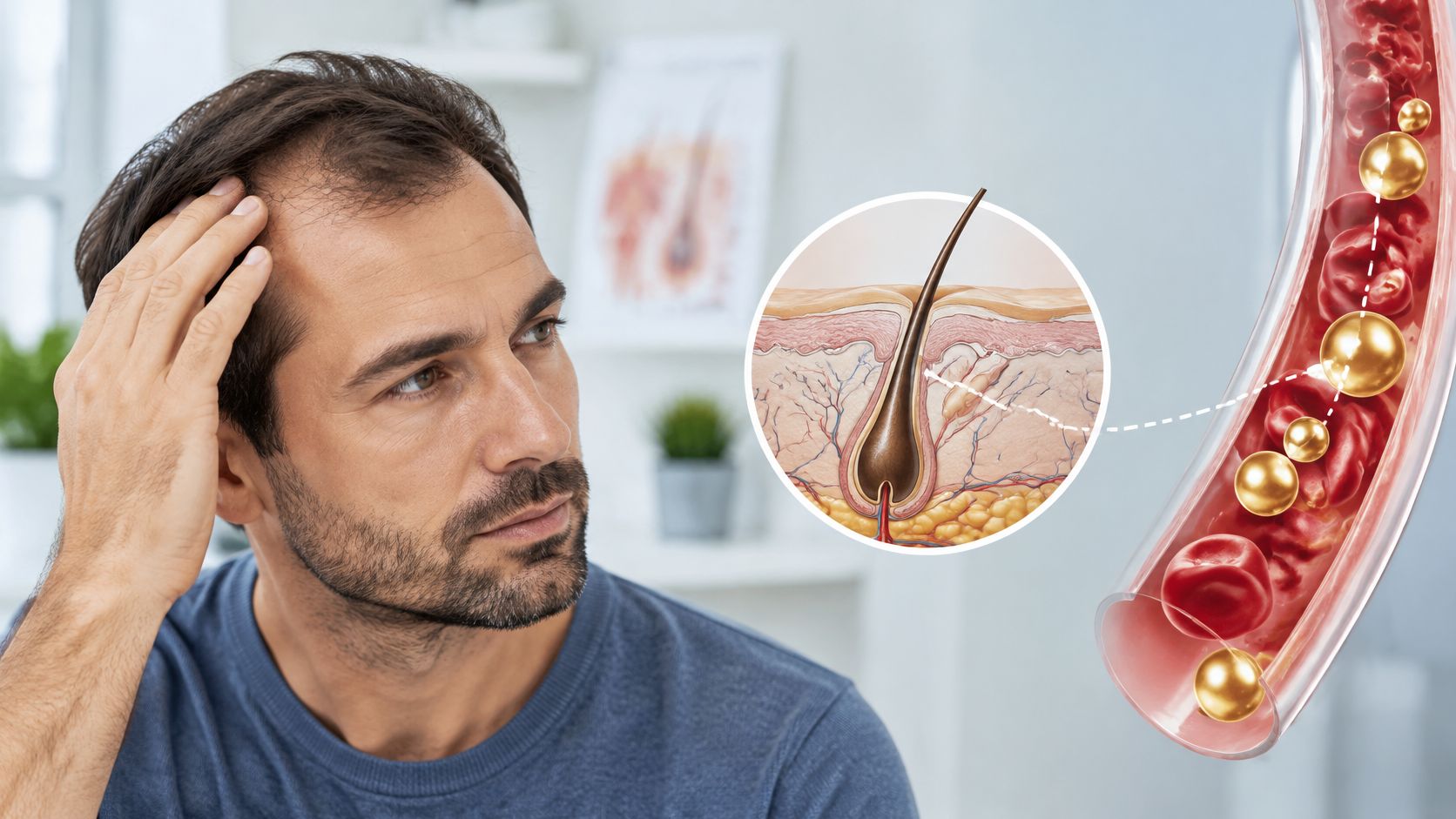
Small Lump on Scalp: Causes and When to See a Doctor
Learn the common causes of a small lump on scalp, which features need prompt medical review, and what to expect at a clinical evaluation.
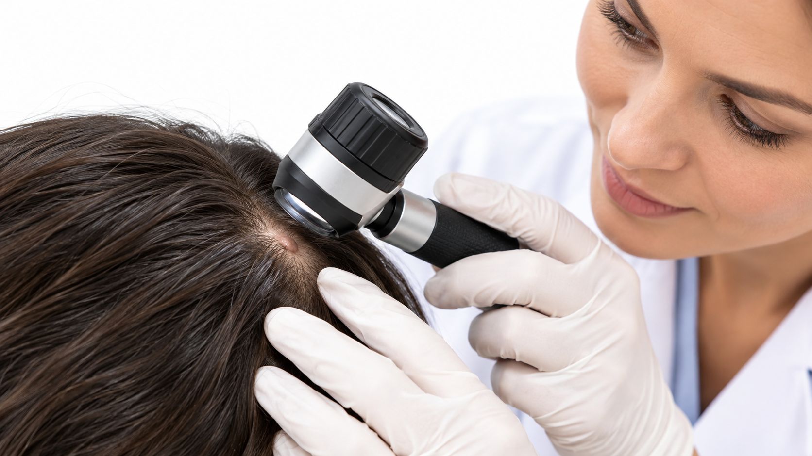
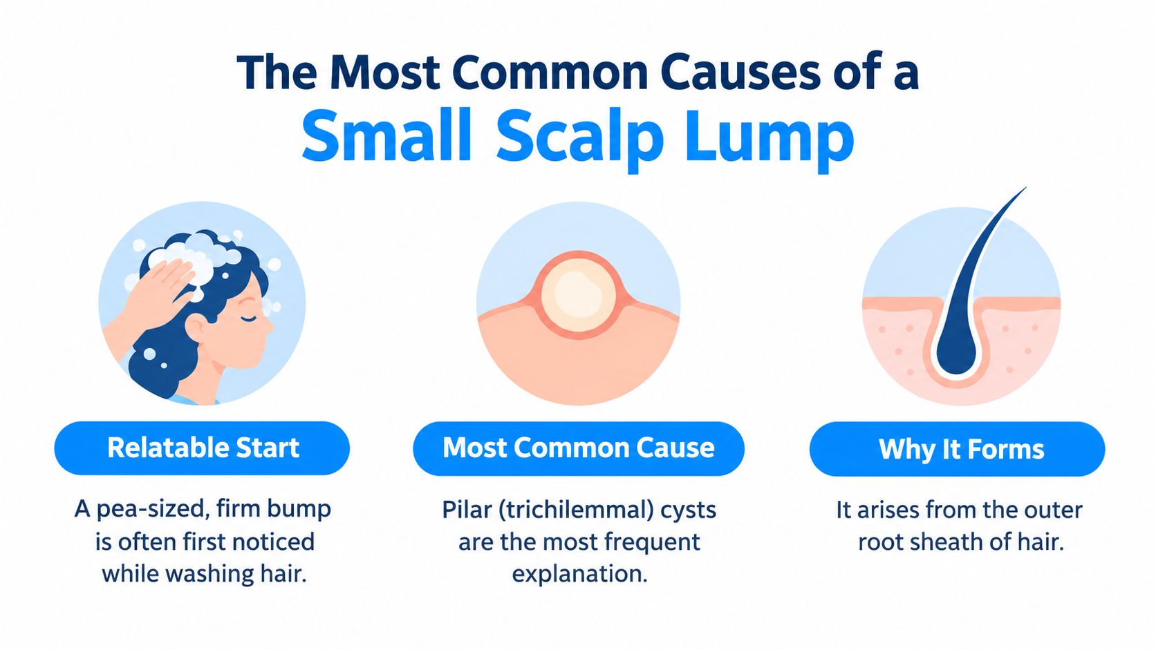
A pea-sized, firm bump discovered while washing hair can raise an immediate question: is it a cyst, an irritated follicle, or something that needs closer attention? Many scalp lumps are benign, but neither a small size nor a painless feel establishes the diagnosis.
The most useful distinction is often how the lump changes over time. A structured record, a clear threshold for assessment, and an appropriate examination are more reliable than comparing the bump with online photographs.
The Most Common Causes of a Small Scalp Lump
Pilar cysts, also called trichilemmal cysts, are among the most common explanations for a small scalp lump. They develop from the outer root sheath of a hair follicle and contain keratin, the structural protein found in skin and hair. Keratin collects within a cyst wall, creating a rounded bump beneath the skin.
According to Radiopaedia's trichilemmal cyst reference, about 90% of pilar cysts occur on the scalp; population estimates range from fewer than 10% of people to approximately 5–10%, while a radiology-based estimate identified them in approximately 2.5% of adults undergoing head imaging. These estimates describe different populations and methods, not the probability that a particular person's bump is a cyst.
What the different lumps can resemble
A pilar cyst is often smooth and slowly enlarging. Some feel firm; others feel fluctuant, meaning that gentle pressure suggests fluid or soft material beneath the surface. Inflammation can make an otherwise quiet cyst tender or swollen.
A punctum is a small central opening in the skin. Pilar cysts usually lack this opening, while epidermoid cysts often have one. That difference can help a clinician narrow the possibilities, but a hidden opening or inflamed surface can make the distinction difficult.
Common scalp lump types compared
| Lump type | Typical feel | Visible punctum | Usual course |
|---|---|---|---|
| Pilar (trichilemmal) cyst | Smooth, firm to fluctuant, mobile | Usually none | Slow-growing, usually painless |
| Epidermoid cyst | Firm, flesh-colored or yellow nodule | Often present; may release keratin | Slow growth; can become inflamed or rupture |
| Lipoma | Soft, doughy, mobile | None | Slow-growing, usually painless |
| Pilomatricoma | Firm; may feel hard from calcification | None | Slow growth; duration is established from the history |
| Proliferating trichilemmal tumor | Can enlarge rapidly and may ulcerate | No reliable distinguishing punctum | Can be locally aggressive; requires clinical evaluation |
Why the comparison isn't a diagnosis
A lipoma consists of fatty tissue rather than keratin. A pilomatricoma is a different hair-follicle-related growth that can become hard because of calcium deposits. These underlying differences aren't always apparent through the skin.
Pilar cysts can also occur in families, so a history of similar lumps among relatives may be relevant. It still doesn't establish what a new lump represents.
The table is a starting point for assessment, not a home identification test. Several conditions overlap in texture and appearance, and an inflamed benign lesion can look more concerning than an uncomplicated one.
Warning Signs That Need a Medical Assessment
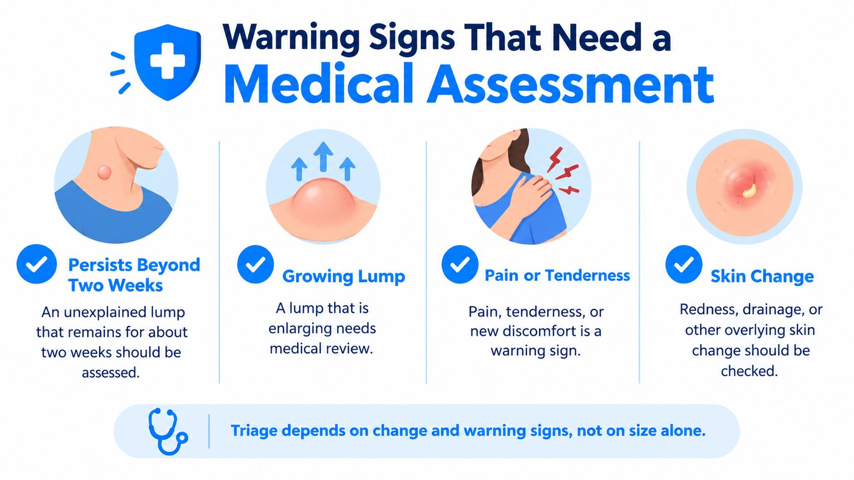
An unexplained lump that persists beyond approximately two weeks should be assessed, according to the NHS guidance on lumps. Assessment should happen sooner when growth, pain, skin changes, or other concerning symptoms develop.
The waiting period isn't permission to ignore an evolving lesion. A small bump that becomes hot and increasingly painful needs a different response from a quiet bump noticed incidentally.
Matching the finding to the response
Scalp lump findings and the action each one calls for
| Finding | Why it matters | Recommended action |
|---|---|---|
| Persists beyond about two weeks | An unexplained lump may need a diagnosis | Book a clinical assessment |
| Enlarges over weeks | Growth can signal a different lesion | Book assessment; keep measurements and photographs |
| Becomes painful, red, or warm | Possible inflammation or infection | Arrange prompt assessment |
| Feels hard and immobile | Fixed or firm masses need examination | Book an in-person assessment |
| Returns after removal | Recurrence changes the possible diagnoses | Book evaluation |
| Bleeding or ulceration | May indicate a more serious lesion | Seek prompt assessment |
| Fever, chills, pus, or spreading redness | Possible abscess or systemic infection | Seek urgent care the same day |
Redness, warmth, escalating tenderness, and drainage can develop when a cyst ruptures or becomes infected. An abscess is a collection of pus that may need treatment beyond home care. Squeezing an already ruptured cyst can spread infection into surrounding tissue.
What a reassuring pattern can and cannot establish
A stable, mobile, non-tender lump without overlying skin changes is more compatible with a benign process. However, those features support a clinical impression rather than exclude every important alternative.
Mobility is especially easy to misinterpret. Hair, scalp thickness, discomfort, and the depth of a lesion can affect how movable it seems under a fingertip. Repeated pressing can also irritate the area and make later comparisons less useful.
Practical rule: The response should follow the most concerning feature, not the most reassuring feature.
Same-day urgent assessment is appropriate for:
- Rapid enlargement: A noticeable, accelerating increase rather than a subtle difference between photographs.
- Severe pain: Particularly when discomfort is worsening or accompanied by swelling.
- Spreading redness: Especially with warmth extending beyond the lump.
- Systemic symptoms: Fever, chills, or feeling generally unwell.
- Pus or significant swelling: Possible signs of an abscess or substantial inflammation.
Telehealth can help organize nonurgent triage, but a photograph can't establish the depth of a mass or reliably assess an abscess. Red flags require an appropriate in-person examination rather than reassurance based solely on a remote image.
Monitoring a Lump Over Time at Home
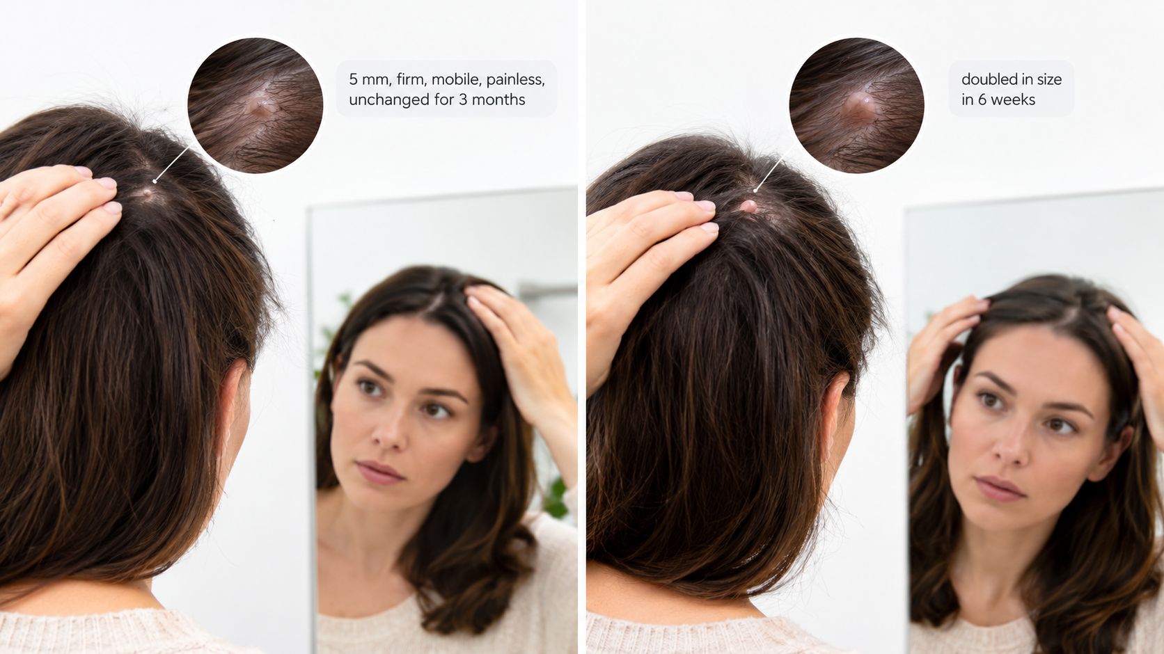
Consider a hypothetical comparison: a 5 mm, firm, mobile, painless lump that hasn't changed since it was found three months earlier, versus a lump that has doubled in size over six weeks. The second pattern calls for assessment because it is enlarging. The first is less concerning in its trajectory, but still deserves an initial diagnosis if it has never been examined.
Home monitoring helps document a lesion; it doesn't replace that diagnosis. The NCBI Bookshelf clinical overview of pilar cysts describes their often asymptomatic presentation and supports tracking persistence and changes rather than diagnosing by touch alone.
Building a useful baseline
A brief written record should include:
- Date first noticed: Including whether the lump appeared suddenly or was discovered accidentally.
- Approximate diameter: Measured with a ruler placed beside the lump, without pressing into it.
- Tenderness: Whether discomfort occurs spontaneously, with brushing, or only when touched.
- Color and surface: Including any visible opening, crust, or broken skin.
- Mobility: A gentle observation, not repeated forceful manipulation.
- Drainage: Its presence, appearance, and whether it occurs without squeezing.
- Photographs: The same hair part, camera angle, lighting, and ruler position for each comparison.
A practical monitoring timeline
At discovery, the record establishes the baseline. At the persistence threshold, an unexplained lump should receive clinical assessment rather than indefinite observation. After a clinician has identified a lesion suitable for monitoring, regular comparisons can follow the interval agreed upon at that appointment.
If a recorded feature changes, the red-flag table determines how quickly reassessment should occur. An uncertain photograph shouldn't delay care when symptoms have clearly worsened.
A painless lump isn't automatically harmless. The absence of discomfort doesn't identify the tissue beneath the skin.
Home squeezing, needle puncture, and attempted excision can introduce bacteria, worsen inflammation, and complicate later removal. A measurement log and clear photographs are more useful contributions to an appointment than repeated attempts to empty the bump.
Common Myths About Small Scalp Lumps
Misleading shortcuts
Small means harmless. Size alone doesn't distinguish a routine cyst from another lesion. A small lump on the scalp still needs assessment when its behavior or examination findings are concerning.
Painless means benign. Many uncomplicated cysts cause no discomfort, but some serious lesions also begin without pain. Comfort isn't a substitute for identification.
Squeezing will solve the problem. Expressing material doesn't necessarily remove the cyst wall. Manipulation can create an inflamed wound while leaving the underlying lesion in place.
Why visual matching falls short
A matching photograph confirms the diagnosis. Similar-looking nodules can arise from different layers and structures of the skin. Lighting, hair coverage, and image angle also hide features a clinician would assess directly.
No change means waiting indefinitely is reasonable. Stability is useful information, but an unexplained persistent lump still warrants an initial assessment. Observation is most useful after a clinician has determined what is being observed.
Only cysts matter. Lipomas, inflamed follicles, injury-related swelling, and enlarged lymph nodes near the back of the head can also produce a noticeable bump. Location and associated symptoms help distinguish these possibilities.
- After an injury: The accompanying symptoms and circumstances matter more than whether the swelling resembles a cyst.
- Near the hairline or behind an ear: A clinician may consider nearby lymph nodes as well as skin lesions.
- With surrounding irritation: Follicular inflammation may be relevant, but deeper swelling still needs appropriate review.
The common error is treating a single clue as a complete answer. A clinician combines the history, physical findings, and any necessary testing rather than relying on a photograph, a fingertip impression, or the absence of pain.
How Clinicians Evaluate Lumps and When Rare Lesions Matter
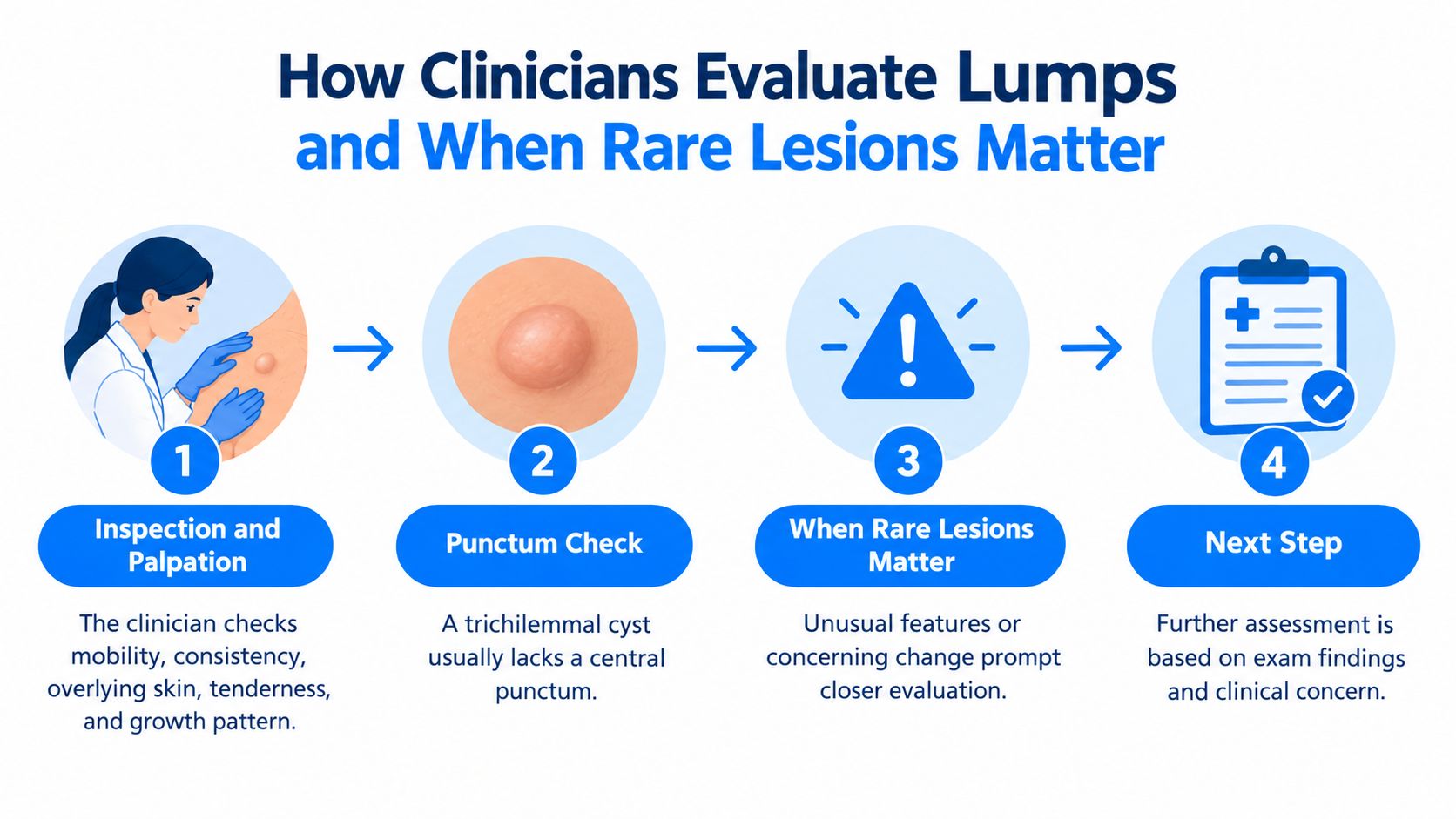
Clinical evaluation begins with questions about onset, growth, prior inflammation, injury, previous removal, and similar lesions elsewhere. Photographs and measurements help distinguish a changing mass from a lump that was noticed recently.
During inspection and palpation, the clinician assesses mobility, consistency, tenderness, depth, and the overlying skin. A pilar cyst usually lacks a central opening, while an epidermoid cyst may release foul-smelling keratin through one, but the DermNet overview of scalp tumors and cysts emphasizes that multiple lesion types can look similar.
Moving from examination to testing
A punctum check means looking for an existing opening. It does not mean puncturing the lesion.
Dermoscopy provides a magnified view of surface structures. Ultrasound can help clarify a lesion's depth and internal characteristics when examination leaves uncertainty. Biopsy provides tissue for microscopic examination when appearance and palpation aren't sufficient.
Evaluation and management by clinical finding
| Clinical finding | Next diagnostic step | Management if indicated |
|---|---|---|
| Stable, mobile, painless, typical cyst | Clinical inspection and palpation | Observation with monitoring |
| Features remain uncertain | Dermoscopy or ultrasound | Plan guided by results |
| Atypical, recurrent, rapidly enlarging, or fixed | Biopsy or imaging | Specialist review; excision as indicated |
| Painful, infected, or repeatedly irritated | Clinical examination | Drainage or excision by a clinician |
| Rupture or abscess | Urgent clinical review | Treatment directed by the examining clinician |
Drainage and excision aren't interchangeable. Drainage addresses collected material, while excision removes the lesion and its wall when appropriate. Active inflammation can affect the timing and type of procedure.
A cyst repeatedly caught by a hairbrush may warrant treatment even without dangerous features. The reason is mechanical irritation, not an assumption that every cyst needs removal.
When uncommon lesions deserve closer attention
A proliferating trichilemmal tumor can resemble a common cyst but may behave aggressively. The Medscape clinical reference on pilar cysts associates this rare variant particularly with older women and a mean presentation age of about 65 years.
A Mexican dermatopathology study published in Dermatologic Surgery in 2023 identified 17 scalp cases across proliferating pilar-tumor and related cyst categories in records spanning 1999 to August 2022, including three malignant cases. This selected case series demonstrates that malignancy can occur; it doesn't establish the cancer risk of an ordinary scalp lump.
Rapid growth, fixation, ulceration, and recurrence warrant closer investigation. Older age adds clinical context, but isn't a diagnosis or an urgent-care criterion by itself.
Tissue examination may be necessary when a lesion's behavior doesn't fit a straightforward cyst.
Hair thinning is a separate clinical concern. An educational guide to hair loss in young men addresses that distinction; medications aimed at hair retention don't identify or treat an unexplained mass. Treatment decisions for the lump belong to the examining clinician.
Choosing Between In-Person Care and Telehealth
Red flags favor in-person care. Rapid growth, bleeding, fixation, significant inflammation, or systemic symptoms require examination at a level of urgency appropriate to the symptoms. A remote appointment shouldn't become an extra step that delays necessary treatment.
For stable findings without concerning skin changes, telehealth may help organize the history and determine whether primary care or dermatology is the appropriate destination. Its value is triage and preparation, not certainty from a camera image.
Choosing the appropriate pathway
- Urgent symptoms: Same-day in-person assessment allows evaluation for infection, abscess, or another rapidly evolving problem.
- A persistent but otherwise quiet lump: A booked examination can establish whether observation is appropriate.
- An already diagnosed lesion under observation: Remote follow-up may support monitoring when the treating clinician considers it suitable.
A useful telehealth intake includes the location, symptom history, previous procedures, relevant medical conditions, photographs, and measurement log. The reviewing clinician should also know whether the lump interferes with brushing, sleeping, or wearing headgear.
Prescription treatments and compounded formulations are prescribed strictly at the clinical discretion of licensed U.S. healthcare providers following an individualized telehealth intake and consultation. An online assessment doesn't establish eligibility or mean that medication is the appropriate response.
Metabolic health, hair retention, sexual wellness, longevity, and hormone protocols address different clinical problems from an unexplained scalp mass. A provider may recommend in-person examination rather than offer treatment through those programs.
Care boundary: Telehealth is useful only when its limitations don't compromise the examination or delay appropriate care.
The strongest next step is the one that resolves diagnostic uncertainty safely. Photographs and a written timeline can improve either type of appointment, but neither substitutes for hands-on assessment when the findings require it.
Vials + Vitals offers physician-directed telehealth assessments for metabolic health, hair retention, sexual wellness, longevity, and hormone care, rather than diagnosis or removal of scalp lumps. Adults seeking support for those separate concerns can visit Vials + Vitals for individualized provider review, while arranging appropriate in-person care for an unexplained scalp lesion.
Informational purposes only. Compounded medications are evaluated by a licensed provider for individual suitability and are fulfilled by state-licensed partner compounding pharmacies.